|
Case of the Month
|
|
CASE 7 & 11 NOT FOR THE SQUEAMISH!
This is a case we just seated yesterday 12/6/01. The photo shown here is in the wax try in stage a few days earlier.
A little history ... This new patient came in with a suspicious area we found immediately on our soft tissue oral exam. The biopsy
showed Dysplasia which I felt had been there already for quite some time. I emphatically explained and implored him to get this
totally removed at once as 100% of these as per Sol Silverman at UCSF (another of my hero figures) will go into full blown cancer.
This patient tried to cure this holistically with herbs ... and sure enough it became malignant. One of our great ENT physicians
(Dr. Kaplan) did a fantastic job on the surgery but had to remove all the maxillary right teeth and bone to the center line
leaving the total area open into the floor of the nose. I had prefabricated an all plastic immediate temporary Obterator
with plastic teeth in place. After healing I had Jamie Moreno at our lab (Golden West Prosthetics) fabricate this swing
lock partial. It fits wonderfully and when locked shut is very stable.
If you will notice I had Jamie leave the inside concave instead of convex so that any food that works it's way up around and
above it, will with with gravity accumulate in the concave portion. This design works wonderfully and looks fantastic!! The
pink plastic covers the metal clasps and actually acts as a clasp its self.
I had had experience early on as the Prosthetic Officer at the Mountain Home Air Force Base in Idaho during Vietnam fabricating
these ... got some fantastic special training at San Diego Naval Hospital. And, as our dental school was very weak in partial
design and fabrication it proved to be invaluable.
Jamie and I have made a lot of these in the past 30 years but most are on the lower.
IT'S NEVER TO LATE TO START LOOKIN FINE! SEE YOUR DENTIST!!
2/10/99
This is just a simple partial denture we made about 15 yrs ago with metal occlusions in the limited distal space = has held up very well
... notice the mesial rests & the I bars .... still no better way to preserve the abutment teeth or more conservative way to make a
standard style partial. On bruxers we make entire dentures out of a Vitalium II metal base with all metal occlusions with the plastic
bonded onto the fronts of the metal where it shows .... they can eat nails with them!!! hee-hee! .... have some that are 28 yrs old
& still going ... some are on their 2nd or 3rd go around of plastic added over the same Vitallium II.
1/26/98 - I THINK THIS MAY BE THE FIRST TIME THIS PROCEDURE HAS EVER BEEN SHOWN! I have never seen it written up before.
This patient fractured off this crown #14 into the tri-furcation area & into 2 separate pieces of root! I couldn't reattach it in any way.
He literally begged me to save it - try anything even if it only holds for a while! Well, this is what I did just today 1/26/98!
On the photos from Left to Right - #1 was how it looked before I cleaned it up. #2 after I cleaned it out. #3 the crown prep with
Hybrid composite build up (I usually use Ti - Core but the hybrid was easier to handle in this tough situation) over 2 kruer posts
tapped, threaded & bonded into the two facial canals. This also showing the extraction site of the extracted lingual root.
Photo #4 is a lingual view - the prep is very parallel.
We covered it with a high noble yellow gold since it went so well. The natural molar was extremely huge to begin with & this crown
will be about the size of a small lower 2nd molar when done & being built on just the two buccal roots. It is ideally situated
directly over the mandibular buccal cusps. This is the same individual we re attached his maxillary left 1st bicuspid a couple of teeth
in front of this one after it had fractured at the gingival or gum area over 4 years ago. I think we have shown #12 on the adhesion page.
(12/9/01 - BOTH THESE ARE STILL IN PLACE!) (11/24/2015 This miracle tooth is till there believe it or not!! I will post a current xray
if I can remember next time Ed is in) Ed has many miracle teeth and every type of modern dentistry there is!
Implants, crowns, bridges, veneers, cerec, etc. still all fixed and non removable.
We also have a similar one that was fractured into 3 separate roots that is now over
20 years old. And, if they fail we would now do the implant option. But posts inside of natural roots are still better than metal
into bone. Now, It is things like this you can't put a stop watch on! Don't let the money managers control your office, time and
life! Take all the time you want on everything - go for the long walk in life, not the sprint! And, eventually develop a practice
with only patients that care enough about their own teeth to maintain them and keep regular appointments !
Below are the finished photos of #12 and #14.
2/10/98 - Remember, these Accucam photos above are mirror image. The upper left photo is tooth # 12 that we re attached at the
gingival level about 4 years ago. The one on the upper right shows # 14 from the lingual with the high noble gold crown in place
over only the buccal roots of #14! The lower left photo shows #14 in occlusion. The LR photo just shows a little of #12 & #14,
neither 12 or 14 are strong enough to support a fixed bridge.
|
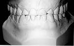
- End to end bite
- Massive abrasion
|
|
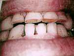
- 13 months of conventional orthodontics
- Extracted 1 lower incisor
- Developed an over jet & overbite
|
|
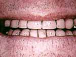
- Only crowned 5 of his teeth
- The rest were bonded where necessary
- Vertical dimension was left unchanged
|
I am particularly proud of this case, as according to the patient, his previous dentist wanted to leave him in his end
to end bite & open his vertical (he had 0 freeway space) and do full mouth crowns for around $40,000.00 None of the posteriors
were in need of crowns as almost all the wear was anterior!

Peg lateral and missing lateral case.
Shaped the left cuspid to look like a lateral & did four porcelain veneers.
Porcelain Veneers, total time from before
to after was 2 weeks.
This is just a boring standard
porcelain to gold bridge with the
laterals turned out, with narrow
& deep embrassures.
HOPE YOU DIDN'T JUST EAT LUNCH!
This benign tumor was removed by
Dr. Rust. It could not have been found
without a Panelipse xray machine.
It is laid out in the position it was
found in the maxillary sinus, at the floor
of the orbit (eye ball). The patient did fine
Kitty cornered - Bi- Cuspid to look like a cuspid,
after making cuspid into
a lateral with a veneer also
Peg Lateral to a full Lateral with a veneer
This site is developed and maintained by C. David Hemp DDS INC
All content Copyright © 1995, 96, 97, 98 by C. David Hemp DDS INC.
All rights reserved worldwide.


